Immunomonitoring

Since the advance of immune-based cancer therapy in the clinic, monitoring the immune status of patients has been increasingly recognized as a critical prognostic parameter. Moreover, prospective clinical and pre-clinical studies are now expected to document immune parameters. In order to respond to this need, the FCI-Immunomonitoring Platform (IMP) has been initiated, with the goal to provide immune monitoring techniques developed by the members of the initiative available to FCI and DKTK researchers and clinicians.

Introduction

The tumor microenvironment and the patient´s immune defense against the tumor play pivotal roles for the progression and outcome of the disease. Paradoxically, tumor-infiltrating immune cells can be anti- and pro-tumorigenic, and the tumor itself can shape the immune milieu. In order to resolve the complexity and heterogeneity of distinct immune cell subsets affecting tumor growth and treatment response in patients with cancer, we have started the Immunomonitoring Platform (IMP) within the framework of the Frankfurt Cancer Institute and the DKTK, where experts have joined to offer a state-of-the-art core unit for researchers and clinicians of the Center.

Flow Cytometry

Technologies

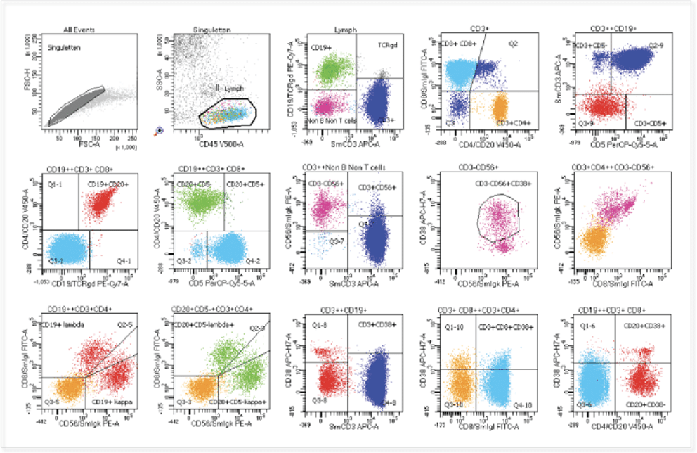
Flow cytometry and Fluorescence-activated cell sorting  (FACS)

Decentralized Flow cytometry and FACS facilities

Workflow: Single cell suspensions are generated with established tissue dissociation protocols, labelled using our antibody panels outlined above, and up to 6 populations are simultaneously FACS-sorted. The sorted cells can then be used in downstream applications.

FACS-Sorting protocols allow isolation of immune cell subsets from human tissue

Focussed and clinically validated multi-color panels to investigate human and murine immune cells in each state of development


  • Blood phenotyping
  • T cell subsets
  • B cell subsets
  • NK cell subsets
  • Lymphocyte differentation
  • Lymphoid diseases
  • Myeloid differentiation
  • Myeloid diseases
  • Monocyte subsets
  • Immune checkpoints
  • Stem / Progenitor cells
  • Neurological diseases
  • Neurovascular unit cells

Exploratory panels (12-35 markers) for immunoprofiling of blood and peripheral tissues

Phenoptics

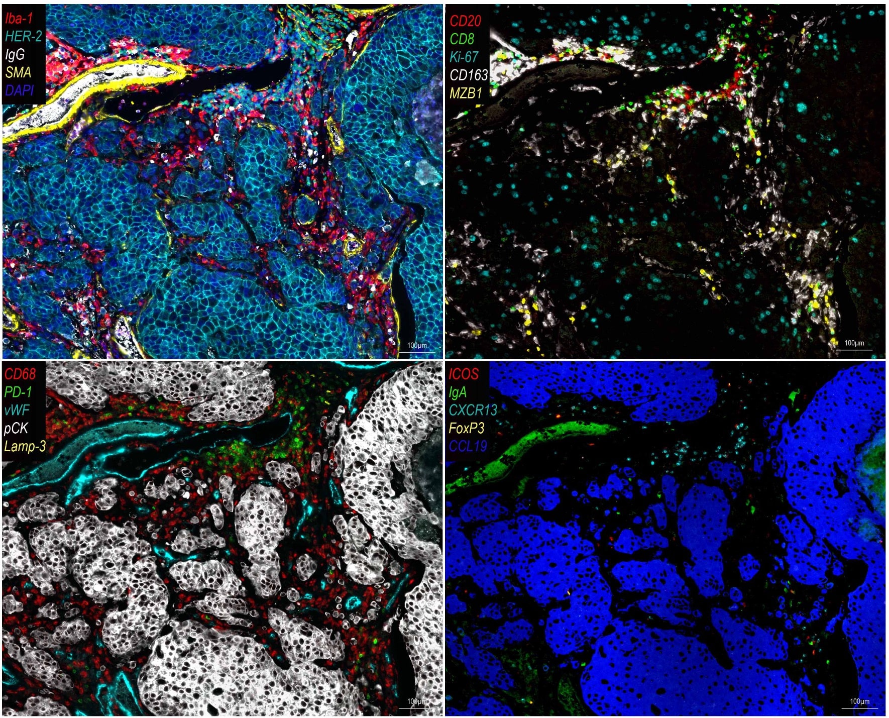
Quantitative spatial information via PhenOptics
(Akoya Biosciences)


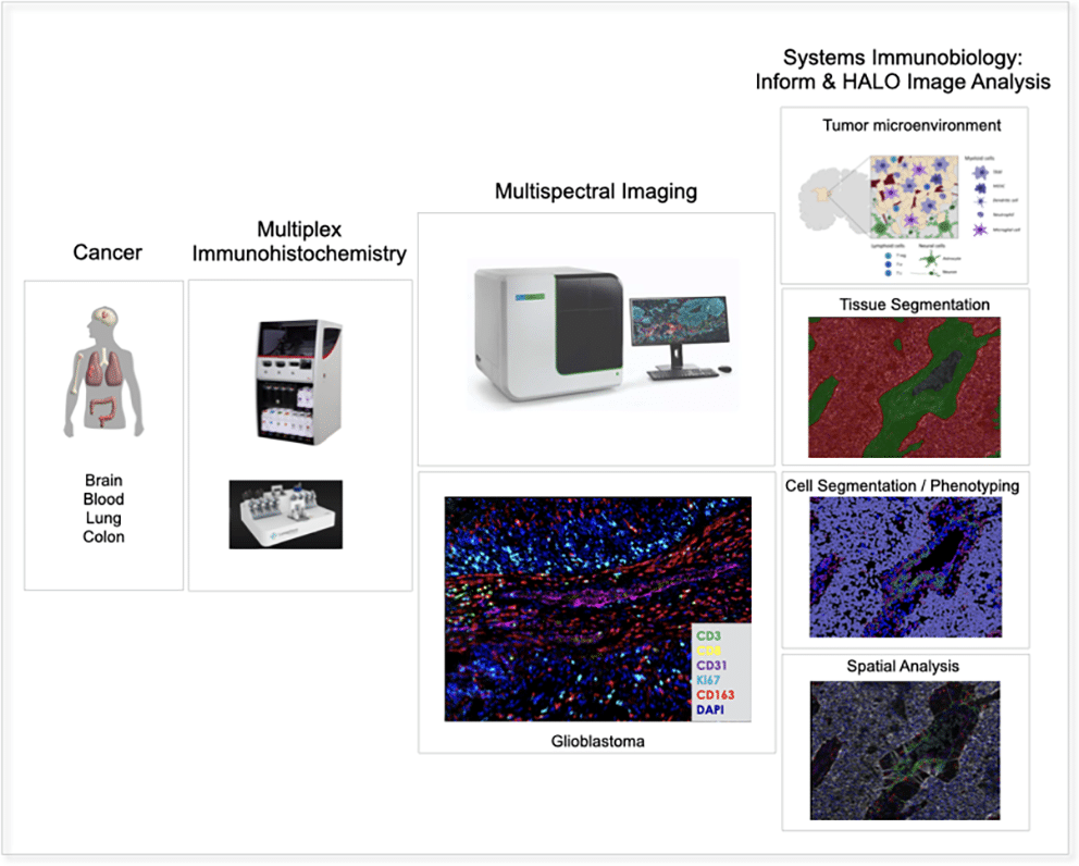
PhenOptics workflowAfter tissue sample acquisition and preparation, sections are stained (7-9 plex) using automated systems (LUNAPHORE LabSat Research Stainer or BOND RX Automated IHC Research Stainer). Multispectral images are generated by the Vectra Polaris Automated Quantitative Pathology System and bioinformatically analyzed by means of inForm Tissue Finder and HALO Image Analysis Software to obtain phenotypic and spatial data.

Single Cell Sequencing

Immune cell multiOmics at single cell resolution

(BD Rhapsody)

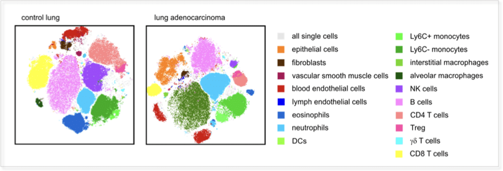
Single cell multiOmics workflow: The BD Rhapsody system combines in a unique way the analysis of gene expression and surface protein expression at a single cell level. Complex heterogenous cell compositions including immune cell subsets in tissue or blood are resolved at highest possible resolution.

Cells are individually paired with beads in the BD Rhapsody, which are coated with barcoded capture oligonucleotides for mRNA. 50-100 oligonucleotide-labeled antibodies can be combined (AB-seq) and will reveal the surface proteome of the cell. The system includes unique molecular identifiers (UMIs) to allow individual transcript counting and can be combined with antibody panels to include protein information in the Bioinformatics pipeline provided within the system. Efficient VDJ- T and B cell receptor sequencing can be applied to study immune cell clonality.

NanoString GeoMx® DSP

Digital Spatial Profiling


NanoString GeoMx® DSP enables researchers to investigate the whole transcriptome or a panel of up to 100 proteins in spatially well defined regions of tissue samples. Barcoded RNA probes or antibodies are incubated with FFPE or fresh frozen tissue samples and later on cleavage via UV light is performed in user defined regions. Readout by NGS on a Illumina NextSeq 1000 sequencer.

Lunaphore COMET™

Highplex Immunofluorescence Staining

To analyze up to 40 proteins at the level of individual cells in a tissue sample, a COMET™ system from Lunaphore Technologies SA is available that automatically stains samples and analyzes them using fluorescence microscopy.

People

The IMP is led by Karl H. Plate, the director of the Edinger Institute and one of the earliest members of FCI.

The IMP members offer a number of state-of-the-art immune-centered assays, share their expertise in experimental design and will help to allocate the most suitable technologies for the required scientific questions.

Please contact us for your future experiments:

IMP Board


Karl H. Plate

Lead

Scientists


  • Franz Rödel
  • Stefan Stein


Staff Scientist:

The Immunomonitoring platform is supported by the

Freunde und Förderer of the Goethe University